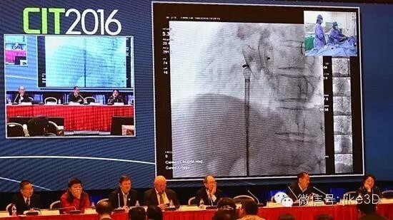
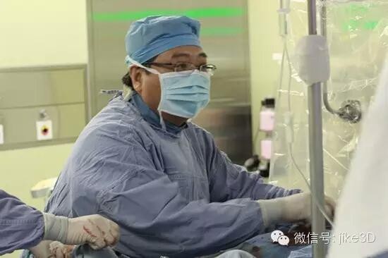
The 14th China Interventional Cardiology Congress was held on March 17-20, 2016 at the Beijing National Convention Center. More than 7,000 experts in the field of cardiovascular disease, interventional radiology and imaging, and other related health fields attended the event. On the morning of March 18th, 2016, I was fortunate to be invited to the cardiac catheterization room of Fuwai Hospital to observe the surgical demonstration and live broadcast of structural heart disease interventional therapy. 2016 CIT International Conference Structural Heart Disease Main Session is undergoing interventional surgery demonstration At 10 o'clock in the morning, the operation demonstration of "interventional treatment of congenital heart disease with multiple atrial septal defect and expanded tumor formation" was officially started by Professor Zheng Hong from the Department of Interventional Therapy of Fuwai Hospital. The patient was treated as a 70-year-old male. A few years ago, he was diagnosed with congenital heart disease due to chest discomfort and his condition was special. Many hospitals were told that surgery was needed. Because the patient is old and fear can not withstand the huge damage of the surgery, he and his family are unwilling to accept and delay until now. This year, the patient's wheezing increased and was recommended by a friend to the Fuwai Hospital for treatment. It happened to meet Professor Zheng Hong's outpatient clinic. His echocardiographic findings were diagnosed as "atrial septal enlargement with multiple atrial septal defect", in which the larger defect was in close proximity to the inferior vena cava. Previously, this inferior vena cava was only treated by surgery. Professor Zheng Hong told his patients that his first use of PDA occluder interventional therapy can solve the problem of not curing the lower cavity type, but suggested that he customize a 3D printed "heart specimen" to understand the "anatomy" of the defect for better. Choose the appropriate size and size of the occlusion device. When it comes to 3D printed cardiac specimens, the heart model, since 2012, Professor Zheng Hong has carried out this research work in China, and has completed the first international “3D printing guided aortic sinus aneurysm rupture intervention†and “3DP technologyâ€. Instructed the use of patent ductus arteriosus occluder for the treatment of inferior atrial septal defect, which also broke the restricted area of ​​the inferior cavity type of insufficiency. So far, Professor Zheng Hong has cooperated with Beijing Instant Medical Imaging Technology Co., Ltd. with independent intellectual property rights to complete the establishment and transformation of thousands of cardiac digital specimen databases and printed nearly 100 specimens of cardiovascular disease. Guided the completion of special or complex cardiovascular interventional and surgical procedures for more than dozens of cases, many of which have been treated with innovations and breakthroughs. Professor Zheng Hong is undergoing interventional therapy for multiple atrial septal defect (central + lower cavity type) in the catheterization room of Fuwai Hospital The ultrasound diagnosis of the interventional treatment case was multiple atrial septal defect, and the large defect was the inferior cavity type. The preoperative MDCT data and the converted 3D cardiac specimen showed three atrial defects and atrial septal enlargement. Tumor, the large atrial defect is narrow and wide, and the lower edge is borderless. At the beginning of the surgery demonstration, Professor Zheng Hong has completed the vascular puncture and right heart catheterization. Due to the expansion of the tumor, bedside ultrasound can not clearly show the shape, location and size of the missing space. Professor Zheng Hong can only use the largest domestic occluder according to the patient's heart "sample" before 3D printing. Blocked. The surgeon first uses the PDA occluder to measure the upper part of the room, and then selects the appropriate size of the occlusion device for sealing. Then the selected PDA occluder is used to seal the lower cavity type. The entire procedure takes about 30 minutes. Although the structure of the case is quite complicated and many experts on the scene during the demonstration broadcast are very different, Professor Zheng Hong successfully completed the operation according to the established plan of his preoperative design. Greenhouse Plastic Plastic Clamps Greenhouse Plastic Plastic Clamps,Greenhouse Structure Connecting Clamps,Agricultural Greenhouses Clamps,Plastic Clamps JIANGSU SKYPLAN GREENHOUSE TECHNOLOGY CO.,LTD , https://www.skyplantgreenhouse.com